
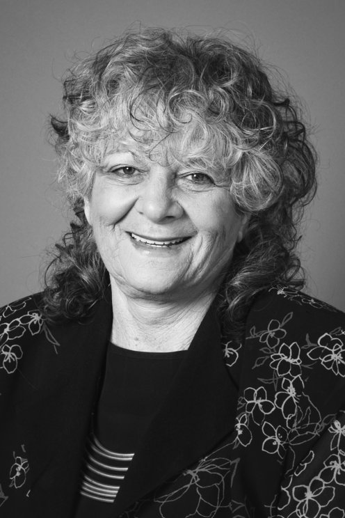
Doctor Ada Yonath is the director of the Helen and Milton A. Kimmelman Center for Biomolecular Structure and Assembly of the Weizmann Institute of Science, a public research university in Rehovot, Israel. She has earned a bachelor’s degree in Chemistry from the Hebrew University in Jerusalem and a Ph.D. in chemistry from the Weizmann Institute of Science. She has held postdoctoral positions at MIT and Carnegie Mellon, and she founded the first protein crystallography laboratory in Israel.1 Dr. Yonath has received numerous honors and awards throughout her career: the Louisa Gross Horwitz Prize for Biology or Biochemistry in 2005, the Paul Ehrlich and Ludwig Darmstaedter Prize in 2007, and the Albert Einstein World Award of Science in 2008, among others. Her most notable prize is the Nobel Prize in Chemistry, which she won in 2009 along with American biochemist Thomas Steitz and Indian-American structural biologist Venkatraman Ramakrishnan.4 Notably, Yonath is only the fourth woman in the world to become a chemistry laureate, and the first Israeli woman ever to win the Nobel Prize.1 She currently lives in Israel with her daughter and granddaughter, working at the Weizmann Institute.5
Background
Ada E. Yonath was born in 1939 in the Geula quarter of Jerusalem. Her father was a rabbi and ran a grocery store with her mother, and despite her family’s poverty, Yonath was able to attend a prestigious secular elementary school called “Beit Hakerem.”8 Interested in science at an early age, Yonath explains that she used to conduct experiments on their apartment balcony: “Once, when I tried to calculate the height of the balcony, I broke my arm. Another time, I wanted to see if water moves faster than kerosene. When my father came out to smoke, a fire broke out.”5 Yonath’s father died when she was 10 and her mother moved with Yonath and her sister to Tel Aviv and took up a job as a treasury clerk. Her family had very little income, so she took up dozens of odd jobs to earn money and support her family: cleaning floor, washing dishes, cleaning her high school’s chemistry lab, and more.3
Yonath spent her compulsory army service in the medical forces, and afterwards, returned to Jerusalem to earn her bachelor’s degree in chemistry in 1962 and her master’s degree in biophysics in 1964 from the Hebrew University. As a graduate student, she attended the Weizmann Institute of Science in Israel and studied X-ray crystallography, receiving her Ph.D. in 1968. During her graduate studies, she studied the structure of collagen, and continued to work on muscle when she accepted a postdoctoral year at Carnegie Mellon.8 After that year, she took up a postdoctoral position at the Massachusetts Institute of Technology (MIT) to study the structure of the protein staphylococcus nuclease.8 It is at MIT that Yonath began investing ribosome structure using X-ray crystallography—research that would eventually win her a Nobel Prize (and is detailed below in Content and Contributions).4
In 1970, after completing her postdoctoral research, Yonath returned to the Weizmann Institute and established the first biological crystallography lab in Israel.8 From 1970-1974 Yonath worked in the chemistry department. In 1974 Yonath became a senior scientist, then an associate professor in 1984, and then the director of the Mazer Center for Structural Biology from 1988-2004.4 Directly after her year as a visiting professor at the University of Chicago (1977-1978), Yonath took on a position as a group leader at the Max Planck Institute for Molecular Genetics in Berlin with Professor H.G. Wittman, who supported her studies academically and financially.8
Throughout this time, Yonath and her colleagues made an astounding 25,000 attempts to get the ribosome to take on a crystalline structure so that it could be easily studied; something that would give scientists unprecedented insight into the mechanisms of gene translation. Finally, in 1980, they succeeded in creating the first ribosome crystals.9 Over the next 20 years, Yonath and her colleagues would continue to improve their techniques and map out the ribosome. In parallel to her activities at the Weizmann Institute, Yonath headed a Max-Planck Institute Research Unit in Hamburg, Germany and made astounding discoveries such as the spatial structure of both subunits of the bacterial ribosome, and how antibiotics can eliminate bacteria by binding to their ribosome and preventing them from translating necessary proteins.9 Her discoveries are vital in understanding antibiotic resistance and creating treatments for many pressing diseases of the 21st century.
Content and Contributions
What is X-ray crystallography? X-ray crystallography is currently the best technique for determining the structure of biological macromolecules. It is used to obtain a three-dimensional molecular structure of a macromolecule from a crystal. This is done when a purified sample of said macromolecule is crystallized at a high concentration and the crystal is then exposed to an x-ray beam. The x-ray beam creates diffraction patterns that can be processed to give information on the symmetry and size of the unit within. The intensities of the pattern can be used to to create a map of electron densities of the unit in the crystal, and the map can be improved until scientists can build a molecular structure from scratch using the protein sequence.10
Yonath began her foray into X-ray crystallography at MIT, when she was studying the structure of collagen. Here, she used X-ray diffraction to study polytripeptides related to collagen and found that some of them give X-ray patterns that are similar to collagen, but not identical. Her results showed that some poly tripeptide structures do not require more than one interchain hydrogen bond per tripeptide.7 Further on, using computers to systematically generate structures with specific bond lengths and angles in order to test their feasibilities, the computations narrowed the possibilities to a conformation which resembles a collagen II model in terms of interchain hydrogen bonding.2
Ribosomal Research
Yonath continued her research into crystallography studying the ribosome. The ribosome was of interest to Yonath because it is incredibly important in human genetics; it is a universal complex that translates genetic code into proteins, and is built by two independent subunits. Ribosomes are also hard to study because they have a natural tendency to deteriorate and have a lot of internal flexibility that can make visualization hard.12
Yonath and her team created the first ribosome crystals in 1980 using a procedure Yonath developed at Weizmann Institute with Professor Ada Zamir, Ruth Miskin, and David Ellison. Taking inspiration from how hibernating bears pack their ribosomes in their cells before hibernation so that the ribosome stays intact and functional for months, they searched for ribosomes from organisms that live under harsh conditions. Eventually, the team used ribosomes from semi-thermophilic bacteria, a hardy bacteria type that lives in extreme environments.8 Thermophilic bacteria that live in hot springs were also studied in order for scientists to extract the DNA polymerase and use it in PCR procedures, since the DNA polymerase of that bacteria could survive incredibly high temperatures. Another procedure Yonath developed is cryo-bio-crystallography, which involves exposing a crystal to temperatures as low as -185°C to minimize structural disintegration under X-rays. After extracting ribosomal information from the bacillus stearothermophilus, Yonath and her team moved onto e. Coli, using similar methods,11 and then more ribosomes.
In the mid-1980s Yonath and her team used their crystallography to visualize a tunnel spanning the large ribosomal subunit: the path through which mRNA progresses as it is being translated into an amino acid. By the mid-1990s, Yonath was not alone in her field any longer. Ribosome crystallography had been proven to be a viable field of study, and several universities and research institutions initiated their own research efforts. By the end of the 1990s, Yonath succeeded in making the first electron density map of the ribosome’s small subunit, and in 2000 and 2001, published the first complete three-dimensional structures of both subunits of the bacterial ribosome.8
An example of one of Yonath’s papers is her paper on the large ribosomal subunit of D50S. She found that the structure of the large ribosomal subunit in D50S, a gram positive mesophilic bacteria, was similar to that of H50S but had a different nucleotide orientation in the peptidyl transferase and in protein structure. She also studied how movements of the L1 stalk could facilitate the exit of tRNA. The large subunit creates the peptide bond in between amino acids and provides a pathway for mRNA. The ribosome of D50S also had a similarity to the ribosome of E. coli and T. thermophilus, which Yonath had previously studied. The large ribosomal subunit has a massive core with two stalks, called the L1 and the L7/12 stalk from it. Its flat surface faces the small ribosome and the round backside faces the solvent used in crystallography. Yonath found that the stalks are flexible, and thus have several possible positions. Furthermore, the RNA fold of H50S and D50S are very similar, but the protein fold shows remarkable differences—D50S even contains proteins that do not exist in H50S. Notably, even similar proteins in D50S show different conformations. Yonath furthermore diagrammed the nascent protein exit tunnel: the tunnel by which mRNA goes through the ribosome and is translated. By comparing the tunnels in H50S and D50S, she found that the tunnel has low polarity and large hydrophobic patches.12

Fig 1. The ribosomal structure of the large ribosomal subunit of D50S bacteria12
Bibliography
- Larson, Erik J., and PhD. “Top Influential Chemists Today | Academic Influence.” academicinfluence.com, April 29, 2020. https://academicinfluence.com/rankings/people/most-influential-chemists-today.
- Yonath, A., and W. Traub. “Polymers of Tripeptides as Collagen Models.” Journal of Molecular Biology 43, no. 3 (August 1969): 461–77. https://doi.org/10.1016/0022-2836(69)90352-0.
- UNESCO. “Ada E. Yonath: ‘the Challenge of Science Is like Climbing Mount Everest.’” UNESCO, January 24, 2018. https://en.unesco.org/courier/2018-1/ada-e-yonath-challenge-science-climbing-mount-everest.
- Rogers, Kara. “Ada Yonath | Israeli Biochemist.” Encyclopedia Britannica, n.d. https://www.britannica.com/biography/Ada-Yonath.
- Siegel, Judy, and Yaakov, Lappin. “Nobel Prize Winner ‘Happy, Shocked.’” The Jerusalem Post | JPost.com, October 7, 2009. https://www.jpost.com/Health-and-Sci-Tech/Nobel-Prize-winner-happy-shocked.
- NobelPrize.org. “The Nobel Prize in Chemistry 2009,” 2020. https://www.nobelprize.org/prizes/chemistry/2009/yonath/facts/.
- Traub, W., and A. Yonath. “Polymers of Tripeptides as Collagen Models.” Journal of Molecular Biology 16, no. 2 (April 1966): 404-IN24. https://doi.org/10.1016/s0022-2836(66)80182-1.
- NobelPrize.org. “The Nobel Prize in Chemistry 2009,” n.d. https://www.nobelprize.org/prizes/chemistry/2009/yonath/biographical/.
- http://www.weizmann.ac.il. “Prof. Ada Yonath,” n.d. https://www.weizmann.ac.il/YonathNobel/.
- Smyth, M S, and J H J Martin. “X Ray Crystallography.” Molecular Pathology 53, no. 1 (February 1, 2000): 8–14. https://doi.org/10.1136/mp.53.1.8.
- Wittmann, H.G., J. Müssig, J. Piefke, H.S. Gewitz, H.R. Rheinberger, and A. Yonath. “Crystallization of Escherichia Coli Ribosomes.” FEBS Letters 146, no. 1 (September 6, 1982): 217–20. https://doi.org/10.1016/0014-5793(82)80739-4.
- Harms, Joerg, Frank Schluenzen, Raz Zarivach, Anat Bashan, Sharon Gat, Ilana Agmon, Heike Bartels, François Franceschi, and Ada Yonath. “High Resolution Structure of the Large Ribosomal Subunit from a Mesophilic Eubacterium.” Cell 107, no. 5 (November 2001): 679–88. https://doi.org/10.1016/s0092-8674(01)00546-3. (https://www.weizmann.ac.il/csb/faculty_pages/Yonath/HarmsCELL2001.pdf)

Leave a comment